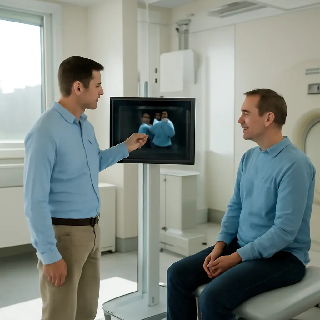
Cardiac imaging combines technologies that show the heart’s structure, movement, blood flow, and tissue characteristics. It is becoming more precise through advances such as cardiac MRI, 3D echocardiography, cardiac CT, and AI-assisted analysis. These tools can help clinicians detect disease, measure risk, and plan treatment more accurately, although the best test depends on the clinical question. The American Heart Association estimates that cardiovascular disease affected 127.9 million U.S. adults in 2020, underscoring the value of timely, appropriate diagnosis.
Cardiac imaging precision: methods, measurements, and clinical value
Precision in cardiac imaging means producing reliable, clinically useful information about a patient’s heart while choosing an appropriate test and limiting avoidable risks. The American College of Cardiology describes cardiovascular imaging as the use of noninvasive and invasive techniques to evaluate cardiac structure and function. In practice, precision depends not just on image resolution, but also on acquisition quality, interpretation, patient context, and whether the result changes care.
Echocardiography: real-time structure and function
Echocardiography uses ultrasound to create moving images of the heart. It is commonly used to assess the valves, chambers, pumping function, and blood flow, and it does not use ionizing radiation. Two-dimensional echocardiography remains widely used; three-dimensional imaging can add spatial detail, while Doppler techniques estimate the direction and speed of blood flow.
A key measure is left ventricular ejection fraction, the percentage of blood expelled from the left ventricle with each contraction. The American Society of Echocardiography’s chamber-quantification guidance recommends standardized measurement methods because image angle, tracing, and image quality can affect results. Strain imaging, which tracks subtle changes in heart-muscle deformation, may reveal dysfunction even when ejection fraction is preserved. These measures are interpreted alongside symptoms and other clinical findings rather than used as stand-alone diagnoses.
Cardiac MRI: tissue characterization and detailed function
Cardiac magnetic resonance imaging (MRI) uses magnetic fields and radio waves to assess cardiac anatomy, function, blood flow, and tissue. It does not use ionizing radiation. MRI can help characterize conditions such as cardiomyopathy, myocarditis, and scar after a heart attack; late gadolinium enhancement, when contrast is appropriate, can make areas of abnormal tissue more visible.
The Society for Cardiovascular Magnetic Resonance publishes standardized protocols to support consistent acquisition and reporting. MRI’s strengths include reproducible ventricular volume measurements and tissue assessment, but access, scan time, patient tolerance, implanted-device compatibility, and the need to evaluate contrast risks can affect whether it is the right test.
Cardiac CT: coronary anatomy and rapid assessment
Cardiac computed tomography (CT) uses X-rays and computer processing to produce cross-sectional images of the heart and coronary arteries. Coronary CT angiography can help assess coronary artery narrowing, while noncontrast CT can measure coronary artery calcium, a marker of calcified plaque burden. CT is also used to evaluate structural anatomy before some procedures.
The 2021 chest-pain guideline from the American College of Cardiology and American Heart Association recognizes coronary CT angiography as an option for selected patients with stable or acute chest pain, depending on risk and clinical circumstances. CT involves ionizing radiation, and angiography generally requires iodinated contrast. Modern scanners and dose-management approaches can reduce exposure, but clinicians still weigh those factors against the expected diagnostic benefit.
Together, ultrasound, MRI, and CT answer different questions: how the heart moves, what its tissue is like, and what its coronary anatomy shows. Newer computational tools build on those modalities by helping clinicians extract and interpret information more consistently.
AI-assisted cardiac imaging: faster analysis and decision support
Artificial intelligence (AI) in cardiac imaging refers to computational methods that identify patterns, measure structures, or assist with image interpretation. The U.S. Food and Drug Administration maintains a public list of AI-enabled medical devices, including authorized products for medical imaging. Authorization does not mean a tool is appropriate for every patient or setting; performance must be considered within its intended use and validated clinical workflow.
Automated measurements and workflow support
AI software can help outline heart chambers, calculate volumes, flag image-quality problems, or prioritize studies for review. Automation may reduce repetitive measurement work and improve consistency, but clinicians must check results, particularly when images are incomplete or a patient’s anatomy differs from the data used to develop the model. A software-generated number is an aid to interpretation, not a diagnosis by itself.
Validation, bias, and clinical oversight
A cardiac AI tool should be tested on data that represent the patients and imaging equipment where it will be used. Important checks include accuracy across demographic groups, performance at different sites, and the effect of image quality or changes in scanning protocols. The FDA’s guidance on AI-enabled device software emphasizes lifecycle management, including monitoring and updates. Hospitals also need clear responsibility for reviewing outputs and escalating uncertain findings.
AI is most useful when it complements established imaging rather than replacing clinical judgment. That principle connects directly to the broader goal of precision: choosing the right modality and interpreting its findings in the context of the whole patient.
Choosing the right cardiac imaging test
The most precise test is not necessarily the newest or most detailed one; it is the test best suited to the patient’s symptoms, risk, and clinical question. The American College of Cardiology and American Heart Association chest-pain guideline recommends a risk-based approach, helping clinicians avoid unnecessary testing while identifying patients who may benefit from imaging.
-
For valve disease, heart failure, or a first look at cardiac function, echocardiography is often an appropriate starting point.
-
For suspected heart-muscle disease, inflammation, or scar, cardiac MRI may provide useful tissue characterization.
-
For selected patients with chest pain or suspected coronary disease, coronary CT angiography can assess coronary anatomy.
-
For suspected coronary plaque burden, a calcium scan may contribute to risk assessment when clinically appropriate.
These are general examples, not individual medical advice. Decisions should account for pregnancy, kidney function, implanted devices, allergies, radiation exposure, availability, and whether the result is likely to alter management. A useful accompanying graphic would compare echocardiography, MRI, and CT by what each measures, radiation use, contrast considerations, and common clinical applications.
What greater imaging precision means for patients
More precise imaging can support earlier recognition of disease, better characterization of risk, and more informed treatment planning. It can also help clinicians avoid repeating tests when a high-quality study has already answered the clinical question. However, additional detail does not automatically improve outcomes: imaging should be ordered when its expected value outweighs its risks, cost, and potential to generate incidental findings.
Patients can take an active role by asking what question a proposed scan is intended to answer, whether it uses radiation or contrast, and how the result could change treatment. They can also share relevant information about kidney disease, pregnancy, allergies, and implanted devices. For clinicians and health systems, priorities include standardized protocols, qualified interpretation, appropriate follow-up, and transparent evaluation of AI tools.
Conclusion
Cardiac imaging is becoming smarter through complementary advances in echocardiography, MRI, CT, and AI-assisted analysis. Each method has distinct strengths: ultrasound provides real-time functional information, MRI offers detailed tissue characterization, and CT can depict coronary anatomy quickly. AI may improve measurement and workflow, provided its outputs are validated and supervised. The broader implication is not simply more imaging, but better-matched imaging that informs decisions responsibly. Patients should discuss the purpose, benefits, and risks of a test with their care team, while healthcare organizations should continue to assess quality, equity, and clinical impact.
Sources: American Heart Association, Cardiovascular Disease Statistics, https://www.heart.org/en/about-us/heart-and-stroke-association-statistics; American College of Cardiology, Cardiovascular Imaging, https://www.acc.org/Guidelines/Guidelines-Resources/Clinical-Toolkits/Imaging; American Society of Echocardiography, Recommendations for Cardiac Chamber Quantification by Echocardiography in Adults, https://www.asecho.org/guideline/chamber-quantification/; Society for Cardiovascular Magnetic Resonance, Standardized Protocols, https://scmr.org/page/Protocols; Gulati et al., 2021 AHA/ACC Guideline for the Evaluation and Diagnosis of Chest Pain, https://doi.org/10.1161/CIR.0000000000001029; U.S. Food and Drug Administration, Artificial Intelligence-Enabled Medical Devices, https://www.fda.gov/medical-devices/software-medical-device-samd/artificial-intelligence-enabled-medical-devices; U.S. Food and Drug Administration, Artificial Intelligence in Software as a Medical Device, https://www.fda.gov/medical-devices/software-medical-device-samd/artificial-intelligence-software-medical-device